Endometrioma
MRI is highly sensitive and specific for the diagnosis of adenomyosis, deep endometriosis, endometriomas, and rectosigmoid endometriosis.
 |
Focal adenomyosis as seen by thickening of the junctional zone, along with punctate high signal foci (dashed arrows). Incidental note is made of Nabothian cysts (arrows). |
 |
Note left ovarian endometrioma (arrow), with a characteristic fluid/fluid level (dashed arrow). |
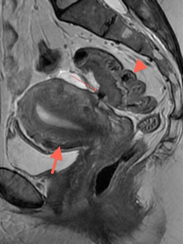 |
Another example of endometriosis, this time with deep endometriosis involving the bowel. Note tethering of bowel (thin arrrow) with bowel wall thickening behind uterus (thick arrow). For comparison note normal thickness of the posterior margin of the bowel (arrowhead |
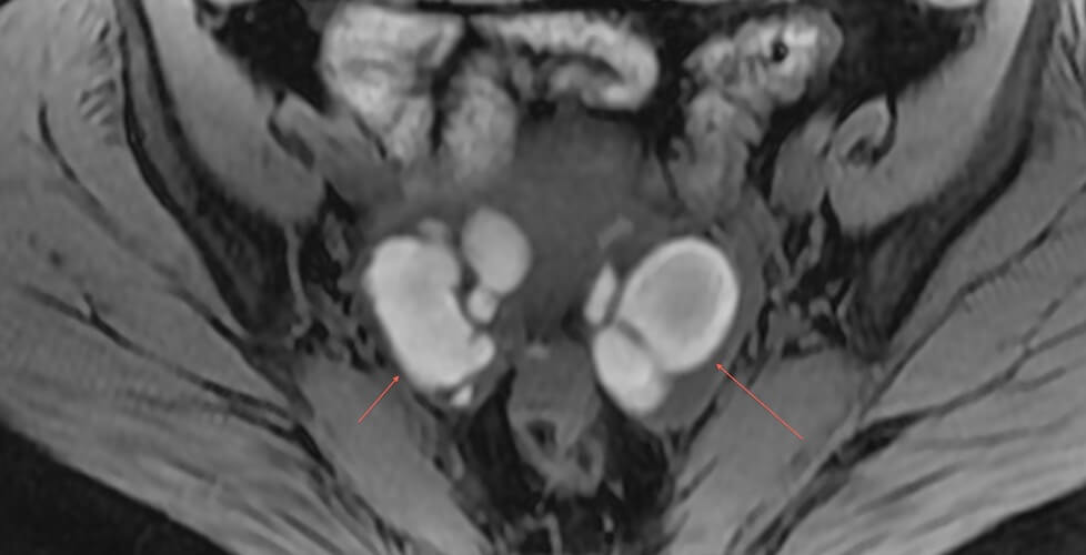 |
Bilateral ovarian endometriomas and right hematosalpinx with bright T1 signal, indicative of hemorrhage. |
References
- Saba L, Guerriero S, Sulcis R, et al. MRI and “tenderness guided” transvaginal ultrasonography in the diagnosis of recto-sigmoid endometriosis. J Magn Reson Imaging 2012; 35:352-360.PubMed link
- Chamie LP, Blasbalg R, Pereira RM, Warmbrand G, Serafini PC. Findings of pelvic endometriosis at transvaginal US, MR imaging, and laparoscopy. Radiographics 2011; 31:E77-100.PubMed link
- Novellas S, Chassang M, Bouaziz J, Delotte J, Toullalan O, Chevallier EP. Anterior pelvic endometriosis: MRI features. Abdom Imaging 2010; 35:742-749.PubMed link
- Marcal L, Nothaft MA, Coelho F, Choi H. Deep pelvic endometriosis: MR imaging. Abdom Imaging 2010; 35:708-715.PubMed link
- Bennett GL, Slywotzky CM, Cantera M, Hecht EM. Unusual manifestations and complications of endometriosis–spectrum of imaging findings: self-assessment module. AJR Am J Roentgenol 2010; 194:S84-88.PubMed link
- Kunz G, Beil D, Huppert P, Noe M, Kissler S, Leyendecker G. Adenomyosis in endometriosis–prevalence and impact on fertility. Evidence from magnetic resonance imaging. Hum Reprod 2005; 20:2309-2316.PubMed link