Accurate imaging is essential for the diagnosis and management of suspected Müllerian anomalies in pediatric patients. We utilize a tailored imaging approach to evaluate these complex conditions. Initial assessment typically includes pelvic ultrasound, and in some cases, 3D ultrasound. When ultrasound findings are inconclusive or detailed anatomical evaluation is required for surgical planning, we proceed with pelvic MRI. Our dedicated pediatric radiologists, with specialized expertise in Müllerian anomalies, provide meticulous interpretation of these studies. Their detailed reports delineate the specific anomaly, assess associated renal anomalies, and guide clinical decision-making. We prioritize a collaborative approach, working closely with pediatric gynecology and urology to ensure optimal patient care. Referring physicians can expect thorough, timely reports and direct access to our team for consultation on complex cases.
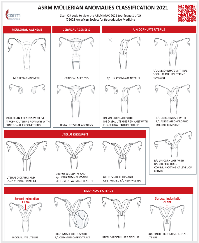
Precise diagnosis classification of Müllerian Anomalies is crucial for determining appropriate clinical interventions and predicting reproductive outcomes. MRI, with its ability to delineate uterine morphology and related structures, serves as the definitive imaging modality. The ASRM classification, as detailed in the following table, provides a systematic framework for interpreting MRI findings, enabling clinicians to effectively manage patients with these congenital anomalies.

Example: Müllerian Agenesis
Example: Uterus Didelphys
Example: Septate Uterus